Pulmonary Valve | Overview
We treat many types of conditions associated with pulmonary valve disease in infants and children. Pulmonary valve disease in the newborn often appears as pulmonary stenosis or a narrowing of the right ventricle. This condition can be treated by our cardiologists in the catheterization lab. If not treated, the right ventricle may not work properly.
Surgical replacement of the pulmonary valve
The most common surgery performed in adults with congenital heart disease is PVR. Although new approaches for less invasive replacement of the pulmonary valve are currently being explored, the standard method is surgical placement of a bioprosthetic valve. Our large experience in PVR has led to a better understanding of the longer-term outcomes after PVR.
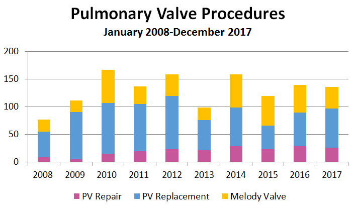
Transcatheter pulmonary valve replacement
Since 2007, we have performed 167 transcatheter PVRs. These procedures are generally performed in our cardiac catheterization lab. Following the procedure, patients are monitored in a short overnight stay and then discharged. Early results have been excellent.
Fetal, neonatal and infant pulmonary atresia, pulmonary stenosis and Tetralogy of Fallot
At the Congenital Heart Valve Center, we focus on minimizing the number of operations children need over a lifetime. For example, we often use catheter-based techniques to treat pulmonary valve atresia and/or stenosis during the fetal or neonatal period. However, there are circumstances in which early surgery is necessary.
For example, TOF occurs in many babies born with congenital heart disease. In these cases, we make every effort to rehabilitate the pulmonary valve. With this in mind, many of our patients can be treated with intraoperative balloon dilation of the pulmonary valve. This approach is intended to alleviate or minimize the need for future pulmonary valve replacements.
Advanced imaging for patients with pulmonary valve disease
Many patients with pulmonary valve disease benefit from advanced imaging developed at Boston Children's Hospital. These technologies provide useful information to help determine the ideal timing for a procedure. For example, MRI can help determined the best time to replace the pulmonary valve.
Pulmonary valve clinical studies and trials
We are currently participating in research and clinical trials seeking to minimize the number of operations children and young adults with disease involving the pulmonary valve (such as Tetralogy of Fallot) need over a lifetime. Many of these patients may also benefit from imaging techniques developed at Boston Children's that provide useful information on the right timing for pulmonary valve procedures.
Pulmonary Valve replacement (PVR)
Patients with TOF often develop severe pulmonary regurgitation. In this case, blood flows backward through the valve, causing an enlarged or “dilated” right ventricle. For these patients, a valve replacement is usually recommended. Over the last few years, we have been working with companies to create a pulmonary valve that can be placed without surgery in the catheter lab.
In 2011, the Food and Drug Administration (FDA) approved the use of a new valve for a certain group of patients. The Medtronic Melody Transcatheter Pulmonary Valve is a prosthetic or artificial device that is implanted through a catheter procedure, rather than a more invasive open-heart surgical procedure. This non-surgical approach makes the child's recovery much easier. Cardiologists at Boston Children’s were among the first to use this valve.
Due to wear and tear, the Melody Valve is likely to need replacement over the course of a patient's life. However, because the Melody Valve can be implanted through a catheter, there is minimal risk to the patient.
Timing for Pulmonary Valve replacement
Repairing the pulmonary valve is not possible for all patients. Many times, a pulmonary valve replacement is needed. Over the last decade, cardiologists at Boston Children's have made a strong effort to identify the optimal time for replacement of the pulmonary valve.
Conditions associated with pulmonary valve disease
- pulmonary stenosis
- pulmonary atresia
- absent pulmonary valve
- with multiple aorto-pulmonary collaterals
Right ventricle to pulmonary artery conduits
- Truncus arteriosis
- Aortic valve disease and Ross procedure
Pulmonary atresaia with intact ventricular septum (PA/IVS)
Endocarditis involving pulmonary valve