Meet Jack
After a prenatal ultrasound, Jack was diagnosed with HLHS. Jack is an example of the innovation of the fetal cardiology team at Boston Children’s.
A fetal cardiac intervention is a highly specialized procedure that Boston Children’s Fetal Cardiology Program first successfully performed more than 20 years ago and has only continued to improve. We created this webpage to explain what a fetal cardiac intervention is, how it’s performed, how we approach it, how your doctor can refer you for an intervention, and what to expect during the procedure.
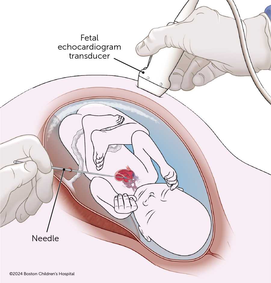
While a fetus is in utero, cardiologists can carefully and precisely help treat a variety of congenital heart defects (CHD) that could otherwise cause serious health problems or may be fatal. A fetal cardiac intervention (FCI) is a needle- and catheter-based procedure guided by an ultrasound image that can help improve some life-threatening heart problems in a fetus.
The first successful fetal cardiac intervention was performed in 2001 by cardiologists and maternal-fetal medicine (MFM) specialists at Boston Children’s and Brigham and Women’s Hospital. Since then, we have advanced the procedure, and our team now offers fetal cardiac interventions for the following conditions:
The procedure starts by the pregnant patient having an epidural for anesthesia and then positioning the fetus in the uterus in a way that the intervention team can access the tiny target in the fetus’ heart. Once optimal positioning is obtained, anesthesia medication is administered to the fetus. The pregnant patient will be awake but able to receive medication to feel more relaxed. They can listen to music on headphones.
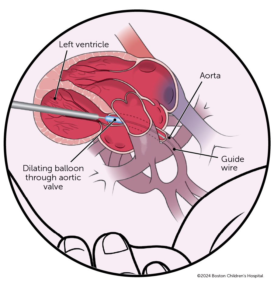
Under the guidance of an ultrasound, our MFM team will insert a long needle, similar to an amniocentesis needle, through the uterus and into the fetal chest and heart. Our interventional cardiologists then thread a balloon catheter through the needle to dilate the narrowed aortic valve, pulmonary valve, or closed atrial septum. The goal of the fetal aortic valve dilation is to open the narrowed aortic valve and increase blood flow — to prevent HLHS and achieve a two-ventricle (pumping chamber) heart, instead of a single-ventricle heart.


The Fetal Cardiology Program at Boston Children’s has performed more fetal cardiac interventions than any hospital in the world. Our team has vast experience and a deep understanding of how to approach cardiac treatments before and after birth.
Our team is comprised of interventional and imaging cardiologists, maternal-fetal medicine specialists, obstetrical radiologists, maternal and fetal anesthesiologists, nurse practitioners, nurses, and social workers. With compassion and an understanding of what your family is experiencing, we work to provide our pregnant patients with the best possible care. Our team has the expertise and experience in treating CHDs before delivery to give children a proper start toward a higher quality of life.
After a prenatal ultrasound, Jack was diagnosed with HLHS. Jack is an example of the innovation of the fetal cardiology team at Boston Children’s.
To determine if you and your fetus are candidates for a fetal cardiac intervention, an in-person evaluation at Boston Children’s Hospital and Brigham and Women’s Hospital is required. The first step of the evaluation, however, is handled remotely; our team will review your fetal echo images while you’re at home. We will consult your primary obstetrical and cardiology team.
If this review determines your fetus is eligible for the procedure, you will then come to Boston for a full day, in-person evaluation to further confirm eligibility for you and your fetus. If eligible, you would then have the procedure.
The morning begins at Boston Children’s:
The next sequence of evaluation steps, mostly at Brigham and Women’s:
If you and your fetus are candidates for a cardiac intervention, the risks and benefits of the procedure will be explained in detail during these consultations. You will need to sign consent forms before proceeding.
The timing of the schedule will depend on the steps of your personalized treatment plan, but the entire process can last between four to six days. Each step below typically transpires over one day, and each step should happen in this order:
Obstetricians and cardiologists, please make early referrals of patients with fetuses showing any signs of aortic stenosis to Boston Children’s Fetal Cardiology Program. Not all fetuses with aortic stenosis will progress to HLHS, but even mild aortic stenosis in mid-gestation can progress rapidly within a couple weeks. The earlier a patient is referred, the more likely a fetal cardiac intervention can be performed in a timely fashion if needed. Please follow the steps below.
Patients, you can also refer yourself. You, too, can follow these steps: