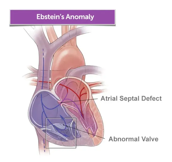
The Congenital Heart Valve Program at Boston Children’s specializes in the care and treatment of tricuspid valve disease. Our team carefully considers two primary approaches when treating tricuspid valve disease: tricuspid valve repair and tricuspid valve replacement. The approach depends on the individual case, as we consider a patient’s condition, the severity of the disease, their heart anatomy, and overall health.
Using innovative three-dimensional modeling — as well as two- and three-dimensional cardiac echocardiography, CT scans, and cardiac magnetic resonance imaging (MRI) — we can see all aspects of a patient’s tricuspid valve disease and their heart anatomy. The perspectives allow us to determine, before surgery, the proper approach to treatment and how we can preserve native tissue. These imaging techniques are particularly important for treating patients who have Ebstein’s anomaly, as they provide us a detailed roadmap that helps us plan that complex repair.
Tricuspid valve repair or reconstruction
Children benefit when their heart valves can be repaired. We’re constantly creating solutions and techniques to repair tricuspid valves so they can remain structurally intact and keep the other parts of the heart strong and healthy. That includes new reconstruction techniques that can improve the function of diseased tricuspid valves that were once considered untreatable. Here are two surgical approaches we take to repair or reconstruct tricuspid valves:
- Cone procedure: Extra tissues on the enlarged right side of the heart are folded, and the malformed tricuspid valve is surgically reshaped into a cone that opens and closes. We typically use the cone procedure to treat Ebstein’s anomaly.
- Starnes procedure: To improve the health of newborns, the tricuspid valve is patched, taking the underdeveloped right ventricle out of the circulation process. This step shifts the workload to the left ventricle, which should eventually form properly with a larger role in blood flow.
Tricuspid valve replacement
Unfortunately, some children have advanced tricuspid valve disease and repairs aren’t enough. They instead need a replacement valve. This involves removing the damaged tricuspid valve and replacing it with a mechanical or biological valve.
- Bioprosthetic aortic valve replacement: Bioprosthetic tissue that replaces an aortic valve is made from animal tissue (pig or cow). A drawback is the animal tissue might not last long and a patient will eventually need another replacement valve.
- Mechanical aortic valve replacement: Mechanical aortic valves are made of strong, durable materials like metal or carbon. They don’t wear out easily and can last a long time. However, the risk of blood clot formation is high, so a patient will have to take blood thinners over a lifetime.
Also, small prosthetic valves are limited in availability, which means young patients may need more interventions as they grow. We are always trying to extend the life of a replacement tricuspid valve and avoid the disadvantages of bioprosthetic and mechanical valves. That’s why we’re constantly searching for new ways to improve the functionality of replacement valves.


