A broken forearm is a break in one (or both) of the bones of the forearm: the radius and/or ulna. The break can occur near the elbow joint, near the wrist joint, or in the middle of the bone. Although the radius can break (fracture) independently of the ulna, it is much more common for the two bones to break together.
Broken forearms are often caused by an impact injury, such as from a fall (as when a child uses his outstretched arm to break a fall) or a direct blow. They are the most common fractures in children, especially if they’re active.
The forearm (lower arm) is the area between the elbow and the wrist. It is made up of two bones: the ulna and the radius.
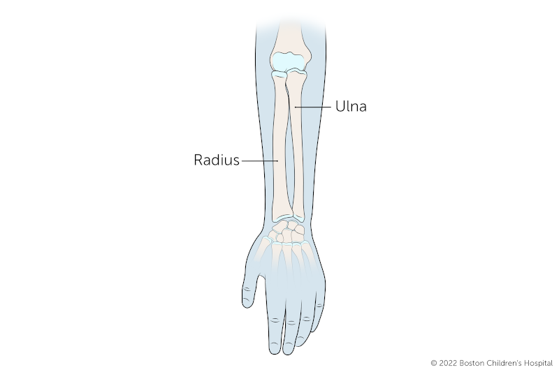
Fractures of the forearm can occur in the radius bone at the wrist, in both the radius and the ulna, and in the ulna alone (near the elbow). Most often, fractures of the radius occur along with injuries to the ulna. Because they are so close together, when one bone is injured, it usually affects the other bone too.
Happens when the radius breaks (fractures) independently from the ulna. When this happens, the end of the ulna can become dislocated at the wrist.
Usually affects the radius or ulna. Children’s growing bones are more elastic than adults’ bones. With excessive force, a child’s bone can experience deformation (bowing) instead of breaking outright. This deformity then remains after the force is removed.
Affects both the ulna and radius. Typically, there’s a fracture of the ulna and a dislocation of the elbow at the top of the radius.
Occurs when the ulna fractures independently of the radius. The ulna can be felt all the way from the tip of the elbow to the wrist, making it particularly vulnerable when children fall to the ground and land on their elbows.
A bone breaks when there’s more force applied to the bone than it can withstand. Childhood broken forearms can be caused by:
To diagnose broken forearms in children, the doctor will carefully examine the injured area for tenderness, redness, and swelling.
One or more of the following imaging techniques may also be used to get detailed pictures of the broken bone and to check for damage to muscles or blood vessels.
An X-ray of the arm is the main tool used for diagnosing a broken bone. This painless test uses small amounts of radiation to produce images of bone onto film. After the doctor puts the pieces of the broken bone in the right position, an X-ray can also help determine whether the bones in the arm are healing in the proper position.
An MRI is a diagnostic procedure that uses a combination of large magnets, radio frequencies, and a computer to produce detailed images of organs and structures within the body. These types of tests are more sensitive than X-rays and can pick up smaller fractures before they get worse.
A CT (or CAT) scan is a diagnostic imaging procedure that uses a combination of X-rays and computer technology to produce cross-sectional images (often called slices), both horizontally and vertically, of the body.
Treatment for a broken forearm depends on the severity of the injury. The goal of treatment is to put pieces of the bone back in place and keep the pieces in the correct position while the bone heals.
Surgery may be needed to put broken bones back into place. A surgeon may insert metal rods or pins located inside the bone (internal fixation) or outside the body (external fixation) to hold bone fragments in place to allow alignment and healing. This is done under general anesthesia.
Splints and casts immobilize the injured bone(s) to promote healing and reduce pain and swelling. They are sometimes put on after surgical procedures to ensure that the bone is protected and in the proper alignment as it begins to heal.
Some common types of casting for broken forearms include:
You can have peace of mind knowing that the skilled experts in our Orthopedics and Sports Medicine Department's Hand and Orthopedic Upper Extremity Program have treated thousands of babies and children with many arm conditions. We provide expert diagnosis, treatment, and care, and we benefit from our advanced clinical and scientific research.
The Orthopedic Clinical Effectiveness Research Center (CERC) at Boston Children's Hospital helps coordinate research and clinical trials to improve the quality of life for children with musculoskeletal disorders. This collaborative clinical research program is unique in the nation and plays an instrumental role in establishing — for the first time — evidence-based standards of care for pediatric orthopedic patients throughout the world.
Major areas of focus for the CERC include:
Ongoing laboratory studies include: