Smith Lab Research | Overview
The Retina and Vascular Development
Our lab investigates factors regulating the vascular development of the retina using several in vivo models. We are dedicated to developing ways to improve therapies for vascular eye diseases, such as retinopathy of prematurity, diabetic retinopathy, and age-related macular degeneration.
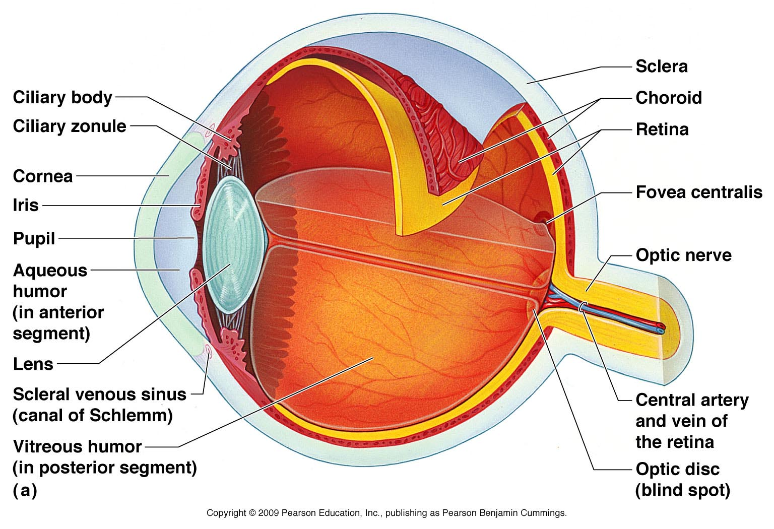
Top: A diagram of the human eye with important structures labeled. Source: Pearson Education, INC.
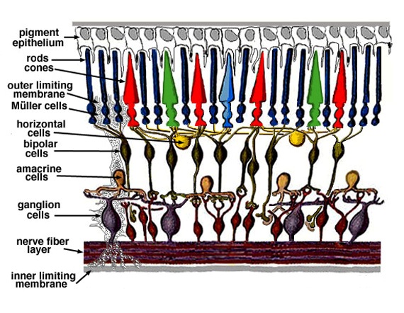
Bottom: A detailed schematic of the layers of the retina. Source: Kolb, Helga. "Simple Anatomy of the Retina by Helga Kolb." – Webvision. N.p., Oct. 2011. Web. 28 Aug. 2015.
A Mouse Model of Vascular Diseases
The mouse model of retinal development has been studied intensively and provides an easily observable representation of vascular development. Unlike humans, mice have an immature retinal vasculature at birth and development continues for the first thirty days. The organized development of the retinal vasculature allows us to reliably observe any abnormal development and investigate the impact of different factors.
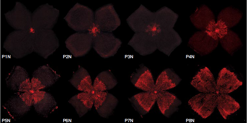
Above: A figure of the vascular development of the mouse retina. Images taken from retinal flatmounts obtained from postnatal day (P) 1 to postnatal day 8. Source: Stahl A, Connor KM, Sapieha P, et al. The mouse retina as an angiogenesis model. Invest Ophthalmol Vis Sci. 2010;51(6):2813-26.
We currently use both transgenic and knockout animals, pharmaceuticals, and dietary modification to investigate multiple genetic pathways regulating retinal development.