Investigating Vascular Eye Diseases
Our lab’s primary research interests are retinal vascular biology in development and in pathological vascular eye diseases, including retinopathy of prematurity, age-related macular degeneration, and rare hereditary diseases such as familial exudative vitreoretinopathy (FEVR) and Norrie disease. We study animal models of these diseases to investigate disease pathogenesis and develop potential therapeutics. Our current research projects are: 1) nuclear receptor-mediated ocular neovascularization and inflammation; 2) small non-coding RNAs in retinal angiogenesis; and 3) Wnt signaling control of vascular growth and permeability in retinopathy.
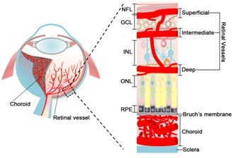
Figure legend: A schematic illustration of the ocular vasculature. Left: A schematic cross-section through an eye showing the retinal vasculature lining the inner surface of the retina and the choroid vessels. Right: An enlarged cross sectional illustration of the eye showing detailed structure of the retinal and choroidal vasculature. Three layers of retinal vessels are embedded among retinal neurons: the superficial retinal vasculature lies in the NFL; the intermediate and deep retinal vascular networks align along each sides of the INL. The choroidal vessels between RPE and sclera serve to supply blood to the outer portion of the retina. GCL: ganglion cell layer; INL: inner nuclear layer; NFL: nerve fiber layer; ONL: outer nuclear layer; RPE: retinal pigment epithelium.