What is an encephalocele?
An encephalocele is a rare birth defect in which the tissue covering the brain, and a portion of the brain itself, protrude through openings in the skull. It may be accompanied by other craniofacial defects.
Encephaloceles can occur anywhere in the skull — involving the top, back, or sides of the head, or the forehead, nose, and eye area. Their severity varies, depending on their size, location, and what parts of head and face are involved.
The exact cause of encephalocele is unknown. It a type of neural tube defect, meaning that the neural tube — a narrow channel that should close during early gestation to form the brain and spinal cord — does not close properly.
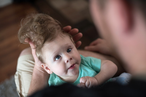
Bentley's second chance
Because of his encephalocele, Bentley wasn't expected to live beyond birth. A one-of-a-kind operation gave him another chance.
Why choose Boston Children’s Hospital for treatment of encephaloceles?
The Craniofacial Program at Boston Children’s brings together a team of highly experienced neurosurgeons, plastic surgeons, oral and maxillofacial (face and jaw) specialists, dentistry professionals, psychologists, and social workers, all experienced in rare malformations. While repairing the physical deformity, we also focus on minimizing disability from any neurologic and developmental damage that may have occurred. Meet our craniofacial team.
Our program has pioneered the use of 3-D printed models of the brain and skull to help plan operations for craniofacial deformities, including encephalocele. The models are created from an individual child’s imaging scans in partnership with Boston Children’s Immersive Design Systems (IDS) Program, allowing surgeons to plan and rehearse complex operations.
Encephaloceles | Symptoms & Causes
What are the symptoms of an encephalocele?
Doctors can see an encephalocele as soon as your baby is born. Sometimes a small encephalocele in the nose and forehead region can go undetected until properly diagnosed. Encephaloceles are often accompanied by craniofacial abnormalities or other brain malformations.
Symptoms of encephalocele that your child may face include:
- neurologic problems
- hydrocephalus: cerebrospinal fluid accumulated in the brain
- spastic quadriplegia: paralysis of the limbs
- microcephaly: an abnormally small head
- ataxia: uncoordinated muscle movement
- developmental delay
- vision problems
- mental and growth retardation
- seizures
Encephalocele can be treated with surgery. Learn more about encephalocele treatment at Boston Children’s.
What causes an encephalocele?
An encephalocele forms when the neural tube does not close properly during gestation. A neural tube is a narrow channel that folds and closes to form the brain and spinal cord. The exact cause, however, is unknown. It usually occurs among families with a history of spina bifida and anencephaly.
Women who are or plan to become pregnant should consume a healthy diet with good sources of vitamin B (folic acid). During pregnancy, an increase in vitamin B may reduce the number of babies born with neural tube defects.
Encephaloceles | Diagnosis & Treatments
How is an encephalocele diagnosed?
Usually encephaloceles are visible deformities and are diagnosed immediately after birth. Occasionally, a small encephalocele in the nasal or forehead region can go undetected.
Sometimes encephaloceles are detected during a routine prenatal ultrasound, at as early as 13 weeks’ gestation. If an encephalocele is suspected on an ultrasound, a fetal MRI can provide all the detail necessary to confirm the diagnosis.
How are encepholoceles treated?
An encephalocele can be treated surgically, ideally during infancy when the skull is still very flexible. The operation involves removing any nonfunctioning brain tissue and cerebrospinal fluid, moving functional brain tissue carefully back into the skull, and relieving any fluid pressure in the head.
The location and size of the encephalocele will determine how much of the defect our surgeons can safely correct. Usually, large protrusions can be removed safely. A complete recovery is most likely when the bulging material consists of primarily cerebrospinal fluid. When a large amount of brain tissue is present in the encephalocele, there is a higher chance of complications during surgery.
Advanced surgery for encephalocele at Boston Children’s Hospital
The largest of its kind in the Northeast, our Cleft and Craniofacial Center offers specialization in rare and complex disorders such as encephalocele. Our team includes some of the world’s most experienced neurosurgeons, plastic surgeons, oral and maxillofacial (face and jaw) specialists, dentistry professionals, psychologists, and social workers — all working together to care for your child.
The Cleft and Craniofacial Center was an early adopter of three-dimensional printing to help surgeons plan for especially tricky cases, making operations shorter and safer. Read more on our science blog and watch how 3-D printing recently guided another type of craniofacial operation.
During surgery, our doctors will:
- Remove the bulging area of tissue, or, if it is living brain tissue, gently ease it back into the skull. This may involve making cuts in the skull bone to make more space.
- Drain excess fluid to relieve pressure inside the skull that can delay normal brain development. Occasionally, shunts (narrow pieces of tubing) are placed to drain excess cerebrospinal fluid from the brain.
The goals of treatment for encephalocele include:
- closing open skin defects to prevent infection and dryness of brain tissue
- removing nonfunctional cerebral tissue outside the brain
- doing a total craniofacial reconstruction
- focusing on minimizing both mental and physical disabilities where neurologic and developmental damage has occurred
Make an appointment
For an appointment with the Cleft and Craniofacial Center, more information or to obtain a second opinion for your child, please call us at 617-355-6309 or email our program coordinator at samantha.hall@childrens.harvard.edu.
International patients
For families residing outside of the United States, please call Boston Children's Global Services at +1-617-355-5209.