What is atrioventricular canal defect?
An atrioventricular canal defect, or AV canal, is a combination of several closely associated heart problems that result in a large defect in the center of the heart. Also known as atrioventricular septal defect or endocardial cushion defect, the condition is congenital, which means it is present at birth, and occurs in two out of every 10,000 newborns. It is often associated with Down syndrome.
When the heart is properly divided, blood from the lungs does not mix with blood from the body; however, with an AV canal, blood moves freely among the four heart chambers. If left untreated, AV canal can cause many problems involving the heart and lungs.
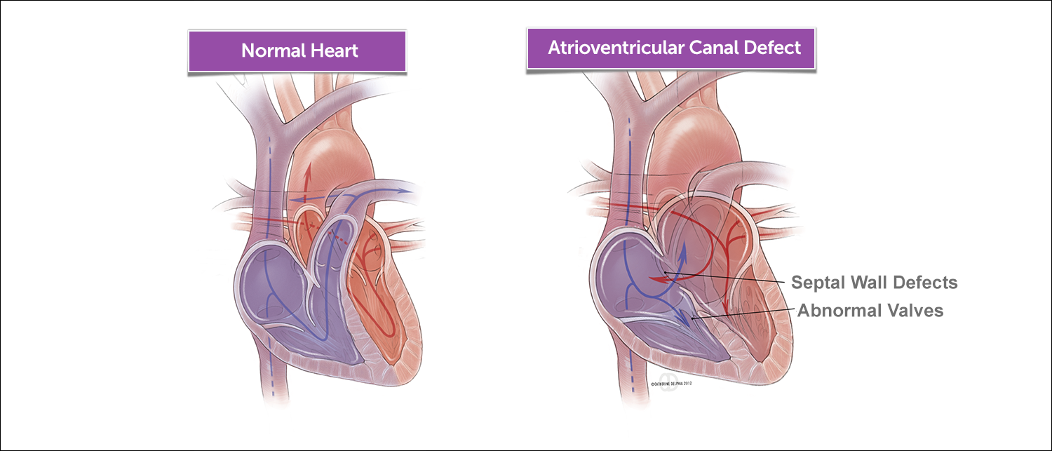
Like many congenital heart conditions, AV canal defect isn’t actually a single defect but rather a group of closely associated defects in various combinations and with varying degrees of severity:
- atrial septal defect
- ventricular septal defect
- abnormalities of the AV valves (mitral and tricuspid) that separate the upper heart chambers (atria) from the lower chambers (ventricles), often resulting in one large “common” valve rather than two separate valves
In addition, there are other associated heart defects such as patent ductus arteriosus, transposition of the great arteries, unbalanced AV canal defects, and aortic arch hypoplasia.
What are the symptoms of atrioventricular canal defect?
A child with atrioventricular canal defect (AV canal) will usually develop symptoms within the first few weeks to months of life. These symptoms may include:
- disinterest in feeding, or tiring while feeding
- poor weight gain
- fatigue
- sweating
- pale skin
- cool skin
- rapid breathing
- heavy breathing
- rapid heart rate
- congested breathing
- blue color
If your child has any of these symptoms, your pediatrician will probably refer you to a pediatric cardiologist for testing, diagnosis, and a determination of treatment.
What causes atrioventricular canal defect?
When the heart is forming during the first eight weeks of fetal development, it begins as a hollow tube. Over time, partitions that form within the tube eventually become the walls dividing the right side of the heart from the left.
Atrial and ventricular septal defects occur when the partitioning process doesn’t occur completely, leaving openings in the atrial and ventricular walls. The valves that separate the upper and lower heart chambers are formed toward the end of this eight-week period, and often they don’t develop properly. Frequently, instead of two separate AV valves (tricuspid and mitral valve), there is a single large common valve that sits between the upper and lower chambers of the heart, allowing blood to flow freely between the chambers above and below the valve — mixing oxygen-rich and oxygen-poor blood.
Do genetics play a role in atrioventricular canal defect?
Genetics may play a role in the development of AV canal defect. Many children have associated genetic syndromes. About 15 to 20 percent of children with Down syndrome also have AV canal defect.
How we care for atrioventricular canal defect
At Boston Children’s Hospital our Cardiac Surgery team treats some of the most complex pediatric heart conditions in the world, with overall success rates exceeding 98 percent — among the highest in the nation among large pediatric cardiac centers. In particular, the methods used to repair an AV canal defect have improved greatly in the past two decades, and the operation has a high likelihood of success.
Our areas of innovation for atrioventricular canal defect
The Boston Children’s Hospital Benderson Family Heart Center deals with the most complex AV canals, which often include children operated on at other centers. We are committed to constantly refining and improving our techniques and procedures.
Children with unbalanced AV canal, traditionally treated as single ventricle defect, are now being considered for biventricular repair. This is achieved using innovative techniques, developed in Boston Children’s Complex Biventricular Repair Program, and refined with 3-D printed hearts in the lab, to ensure the repair is durable throughout a lifetime.
Atrioventricular Canal Defect | Diagnosis & Treatments
How is atrioventricular canal defect diagnosed?
If your newborn has any symptoms that suggest a congenital heart defect, your pediatrician will refer you to a pediatric cardiologist. The cardiologist will perform a physical examination and listen for a heart murmur. The location in the chest where the murmur is heard, as well as the sound and character of the murmur itself, will give the cardiologist an initial idea of what kind of heart problem your baby may have.
What tests are used to diagnosis atrioventricular canal defect?
Some of the following medical tests will be used to diagnose AV canal and its associated defects:
- Electrocardiogram (EKG or ECG) is usually the first test used to diagnose AV canal.
- Echocardiogram is also commonly used for diagnosis.
- Cardiac magnetic resonance imaging (cardiac MRI) is sometimes used in special cases for diagnosing AV canal and may also be used to help plan surgery.
- Cardiac catheterization is only used in rare cases to diagnose AV canal.
How is atrioventricular canal defect treated?
Specific treatments for atrioventricular canal defect (AV canal) depend on the extent of the disease — which can range from a single defect to a full combination of defects (complete). AV canal is almost always treated by surgical repair of the defects. Medications may be helpful and improve symptoms until the operation is performed.
Most children with complete AV canal undergo surgery by the age of 3 to 6 months. Children with partial and transitional AV canal undergo surgery later — 1 to 2 years old. Children with Down syndrome may develop symptoms earlier than other children and may need to have surgery at an earlier age.
Treatments may include:
- Medical management of infants who may become tired when feeding, and may not be able to eat enough to gain weight (Nutritional support from more concentrated breast milk or formula gives the baby more calories or other forms of nutritional-assistance diuretics, such as Lasix, help the kidneys remove excess fluid from the lungs and body.)
- ACE (angiotensin-converting enzyme) inhibitors, such as Captopril or Enalapril to help the heart pump blood forward into the body digoxin and strengthen the heart muscle, enabling it to pump more efficiently
- Surgery to repair the AV canal involving the following components: first, to create two separate functioning AV valves, one for each side of the heart; and second, to close the various septal defects (ventricular and atrial) and address any other additional defects
- 3-D echocardiography to help optimize surgical treatment of the defect to help prolong durability of the valve repair
Care after AV canal surgery
After your baby's operation and hospital stay (usually five to seven days), he or she will need to be followed by a pediatric cardiologist, who will offer recommendations for post-operative follow-up care, including:
- wound care while your baby is healing
- a nutritional program to encourage weight gain
- an oral hygiene program to prevent infection
- an appropriate exercise regimen to build body mass and achieve fitness
As your baby recovers and grows, be sure to follow a regular program of well-baby/well-child checkups.
What is the long-term outlook for atrioventricular canal defect
Many children who've had an AV-canal repair will live healthy lives. Activity levels, appetite, and growth typically return to normal in most children. Some children will still have some degree of mitral- or tricuspid-valve abnormality or leakage after surgery, which may require another operation in the future. Children with AV canal will need lifelong monitoring (some will need medication), since they will always be at some risk for arrhythmias, infections, heart failure, or stroke.
Your cardiologist will help you create a long-term care program as your baby matures into childhood, the teen years, and even adulthood. Most people who’ve had congenital heart disease repair will have an ongoing relationship with their cardiologist. We will prevent and treat complications and will advise on daily life issues, such as activity levels, nutrition, and any precautions related to becoming pregnant.